
The first ultrasound in pregnancy is often an early dating scan performed prior to 10 weeks in gestation. This scan is commonly performed at 7 weeks gestation.
Why scan at 7 weeks?
At approximately 7 weeks gestation, a pregnancy sac growing inside the uterus can be seen. Within that sac, an embryo with a beating heart can be identified. Twins or triplets can also be identified at this stage. Prior to 7 weeks, it may be too early to see the embryo and the heartbeat.
If the pregnancy has occurred through IVF, the 7 week scan is performed approximately 5 weeks after the embryo transfer.
How is the scan performed?
To obtain the best images possible, this scan is routinely performed internally (transvaginal). An internal ultrasound is perfectly safe to perform during pregnancy.
What can be seen during the ultrasound?
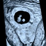
During an early dating ultrasound, the pregnancy sac and embryo will be measured. The length of the embryo helps to determine or confirm the due date of the pregnancy. The heartbeat of the embryo can be measured. The maternal ovaries and cervix are also examined.
What if I have an uncertain result?
On occasion, it may be too early to gather all the information required to confirm the stage of the pregnancy. In this situation, a repeat ultrasound may be necessary to check on the development and re-measure the pregnancy sac and embryo. The referring doctor will determine the most appropriate time frame to return for this scan if required.