What is a pelvic ultrasound?
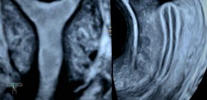
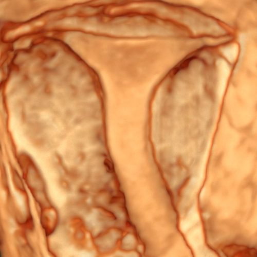
A pelvic ultrasound examines the uterus, ovaries, cervix and other surrounding structures. Ideally and where appropriate, the pelvic organs are best assessed through an internal pelvic ultrasound (transvaginal ultrasound). Alternatively, a pelvic ultrasound can be performed through the lower abdomen (transabdominal ultrasound).
What is an internal (transvaginal) ultrasound?
An internal ultrasound involves a specially designed, slender ultrasound probe being inserted into the vagina and moved gently in order to visualise and examine the pelvic organs. The ultrasound probe is covered in a sterile cover with lubricant gel.
The advantage of the internal approach is the close proximity to the pelvic organs. This allows for the clearest view and most accurate results.
At times during the scan the sonographer or doctor may also apply gentle pressure to the lower pelvis to help visualise the organs.
At any time during the scan, the patient has the option to cease the internal examination if there is pain or the patient is no longer comfortable to continue.
An empty bladder is required for an internal ultrasound.
What is an external (transabdominal) ultrasound?
An external ultrasound involves the ultrasound probe being moved across the lower pelvis to examine the pelvic organs.
The internal ultrasound produces clearer images in comparison to the external ultrasound. The external approach is however the appropriate scan type for underage patients, those who have not previously been sexually active or those patients who do not consent to an internal examination.
A full bladder on arrival for the appointment is an essential requirement of an external ultrasound.
Why is the ultrasound performed?
There are many reasons a patient may be referred for a pelvic ultrasound.
This can include:
- General check up
- Family history of cancer
- Abnormal bleeding
- Pain
- Fertility check
It may also be performed to monitor or examine for the following conditions:
- Fibroids
- Polyps
- Cysts
- Endometriosis/Adenomyosis
- Hydrosalpinx
Who performs the pelvic ultrasound?
The ultrasound is performed by a female sonographer. There are male and female doctors (obstetrician/gynaecologist/sonologists) who may also scan on occasion.
When can I book a pelvic ultrasound?
A pelvic ultrasound can be performed at any time however often the clearest images are obtained immediately after the period and prior to ovulation (approximately between day 7 and day 12 of a 28 day cycle). A pelvic ultrasound is safe during pregnancy and can still be performed where there is spotting or bleeding if unavoidable.
How long do the results take?
A reporting doctor (sonologist) reviews the images after each scan and a written report is forwarded to the referring doctor thereafter.