A routine ultrasound examination is offered to patients at 21 weeks in pregnancy.
The purpose of this examination is to assess:
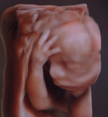
- Fetal anatomy – including a detailed examination of the baby’s brain, face, spine, heart, abdomen, limbs and gender.
- Position of the placenta.
- Amniotic fluid volume.
- Pelvic anatomy and cervix.
This ultrasound is often referred to as the morphology scan. The examination is expected to detect major fetal malformations if present. Ultrasound cannot detect all abnormalities. Some abnormalities are progressive and unable to be detected at this stage. Other anomalies including cerebral palsy, biochemical abnormalities and some chromosomal abnormalities can also not be detected.
The ultrasound may be limited by the following:
- Fetal position
- Increased distance between the fetus and the ultrasound probe. This may occur in the case of very large fibroids or larger amounts of fatty tissue.
Every effort is made at the time of the appointment to ensure that all the required images are taken and are of high quality. There may be times however when a repeat scan is indicated to achieve adequate images.
Please inform the sonographer at the beginning of the ultrasound if the gender of the baby is to remain unrevealed.